
The software automatically suggests all relevant landmarks. These can be selected with a left mouse click – click and hold the left mouse button to move the points. The maxillary tuberosity and the incisive papilla are outlined with a closed line. The centre point (orange) marks the “highest” point of the outlined area and changes its position accordingly when the boundary line is changed.
The green points mark the labial frenum, buccal frena, and the position of the pterygomandibular raphe.
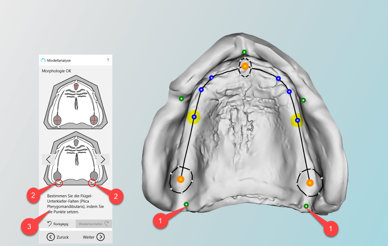
The wizard (left field in the main screen) guides you through the task step by step (3). If an item is selected (1), the corresponding explanation of the step appears in the wizard’s window (2). The sequence of adjusting the points can be done in any order.
The other points on the alveolar process (blue) mark anatomically relevant points:
- Position of the canines – in extension of the first major palatal fold on the alveolar process. If the palatal folds are difficult to see, scarring on the jaw can also give an indication of this position, since canines are usually the longest teeth remaining in the jaw – and are therefore the last teeth to be lost.
- Approx. 3,5 – 4 mm behind the position of the canines, the position of the first upper premolars can be determined. This position forms in connection with the centre of the maxillary tuberosity the line of the basic statics for each side. Along this line the largest elevation of the bone structure of the alveolar process of the upper jaw can be found. The line of the basic static is automatically suggested by the software in the next step.
- Transfer of the deepest point in the lower jaw (masticatory center) to the upper jaw.